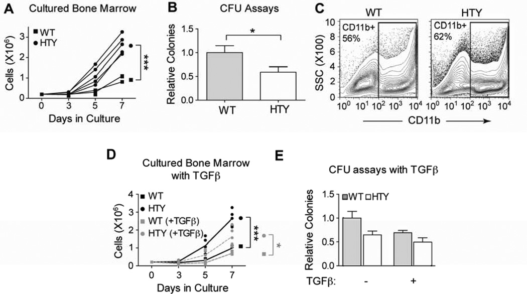
Figure 2. Increased ex vivo growth and diminished colony forming ability of Runx1HTY350-352AAA marrow.
(A) Ex vivo cultures of bone marrow cells from wildtype or Runx1HTY350-352AAA animals (each point represents the mean cell number of technical replicates from one animal; n=3 WT, 5 HTY). (B) Myeloid colony forming unit assays performed with wildtype or Runx1HTY350-352AAA bone marrow cells (n=28 WT, 34 HTY). Because there was no discernable difference in colony size, only total colony number was counted. (C) Cells from the experiment reported in panel A were harvested at day 7 and the percentage of cells expressing the myeloid antigen CD11b was determined using flow cytometry (n=3 WT, 3 HTY). (D) Ex vivo cultures as in panel A, but with the addition of TGF beta to a final concentration of 10ng per ml (each point represents technical replicates from one animal; n=3 WT, 5 HTY). (E) Colony forming unit assays were performed as in panel B, but with the addition of TGF beta to some cultures to a final concentration of 10ng per ml (n=3 WT, 5 HTY). * p<0.05, *** p<0.001, calculated by Student’s T test. All error bars are SEM.

