Figure 7.
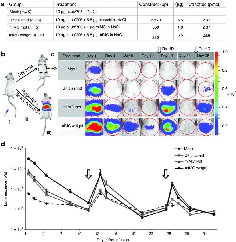
Splice correction from micro-minicircles (miMCs) and plasmid in vivo using the pCMVLuc/705 reporter construct. (a) Experimental setup for the different treatment groups. (b) Schematic presentation of the in vivo procedure with (i) hydrodynamic infusion to the liver of either (ii) pCMVLuc/705 + U7-splicing construct (miMC or plasmid) or pCMVLuc/705 + “mock.” When the luciferase pre-mRNA is correctly processed, the luciferase protein together with its substrate (luciferin) will emit light and the luminescence can be (iii) detected and measured in the IVIS. (c) IVIS-images of luminescence of the mouse liver over time for the four different groups. Each row depicts the same representative mouse for each group measured at the indicated time-points. The red color indicates a stronger signal and blue indicates a weaker. At day 0, all animals received a coinfusion (hydrodynamic infusion to the liver) with pCMVLuc/705 + splicing agent (miMC or plasmid) or mock. At day 12 and 24, reinfusion was made with pCMVLuc/705 to determine the residual activity of the splicing agent (MC or pU7). (d) Graph presenting the mean values of luminescence at log scale for each group of mice over time. Reinfusions of pCMVLuc/705 are indicated by arrows. Data shown as mean + SEM.

