
Fig. 3.
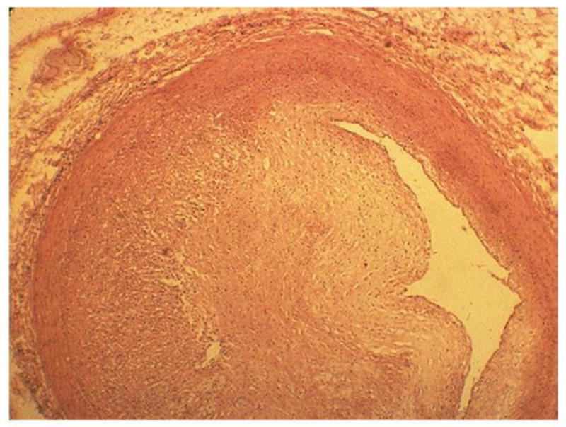
Left anterior descending coronary artery in a 16-year-old boy 1 year after receiving 40 Gy mantle radiotherapy for Hodgkin’s disease. Myointimal proliferation has considerably narrowed the lumen. Fatal cases like this in a patient who had no cardiac risk factors other than radiation illustrate that the morphology of arterial disease due to radiation is essentially no different from that of age-related atherosclerosis. Hematoxylin-eosin stain. (From Fajardo LF. The pathology of ionizing radiation as defined by morphologic patterns. Keynote lecture. 5th Nordic Conference on Radiation Oncology, Bergen. Norway. Acta Oncologica 2005;44:13–22; with permission.)