
Fig. 6.
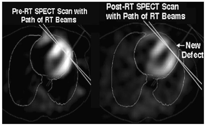
Representative axial images pre-RT (left panel) and post-RT (right panel) cardiac SPECT scans. The deep borders of the tangential RT beams are shown as solid lines. A new perfusion defect in the anterior left ventricle after radiation is seen.