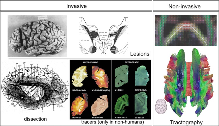
Figure 1.

Available techniques for measuring anatomical connections in the brain. Lesion studies rely on Wallerian degeneration as a result of a brain lesion; the effects of the lesion can be seen postmortem at remote sites (here the thalamus) indicating the trajectories of white-matter projections (from Ref. 98). Postmortem dissections of white-matter connections date back to the 19th century (from Ref. 99). A multitude of tracers are available in animals. Shown here are example anterograde (biotinylated dextran amines, or BDA) and retrograde (Fast Blue fluorescent dye) tracers used to trace connections from the posterior cingulate cortex in macaques (from Ref. 100). The only available technique that is noninvasive is diffusion MRI tractography. The panel on the right shows how local estimates of fiber orientation, here using the diffusion tensor model, can serve to trace estimates of neural pathways. This allows reconstruction of major white-matter connections in the whole brain (top: figure from Ref. 31; bottom: image courtesy of Alexander Leemans).
