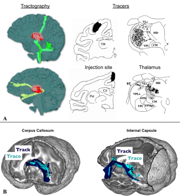
Figure 2.

Two example comparisons between tractography and tracer results. (A) Connections traced from two locations in the thalamus using human dMRI tractography (left-hand side, modified from Ref. 74). Tracer studies in monkeys (right-hand side, modified from Ref. 101) shows that different thalamic regions contain traces of the injected dye depending on the cortical injection site. Comparing the two allows us to interpret the tractography result in terms of the location of the tractography seed relative to different thalamic nuclei. (B) Direct comparison of tractography and tracing of the same connections in the macaque brain. Shown here are two connections from the lateral orbitofrontal cortex traveling through the corpus callosum and the internal capsule, respectively, with a very good match between the two techniques. Modified from Ref. 47.
