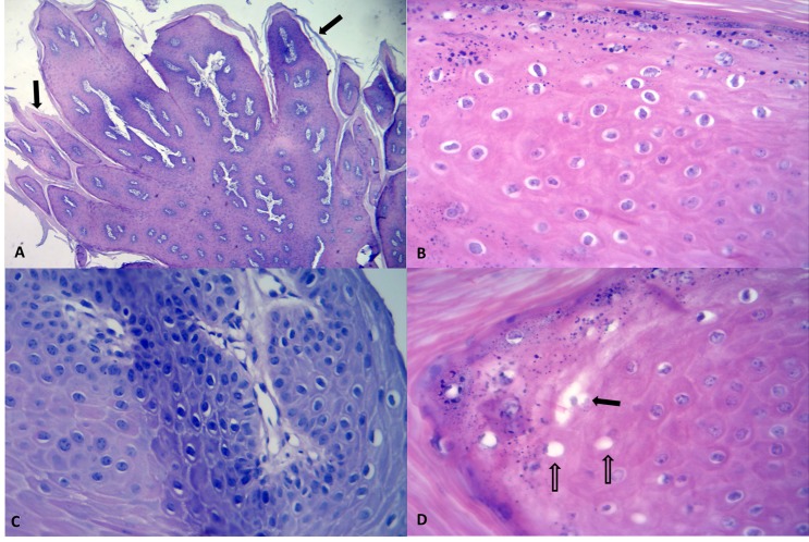
Figure 1.
Histopathological features of teat papillomas; skin, mammary gland, cow. There is outward digital-like proliferation of the squamous epithelium with hyperkeratosis (arrows) of the stratum corneum (A). Observe that the nucleuses of most keratinocytes are surrounded by a clear halo (B) and the severely condensed basophilic-staining (pyknotic) nucleus (C) of the koilocytes. There are several degenerated keratinocytes (open arrows) and observe some that adjacent swollen cells are coalesced into a larger microvescile (closed arrow) (D). (A, Hematoxylin and eosin, 4 × Obj.; B–D, Hematoxylin and eosin, 40 × Obj.).

