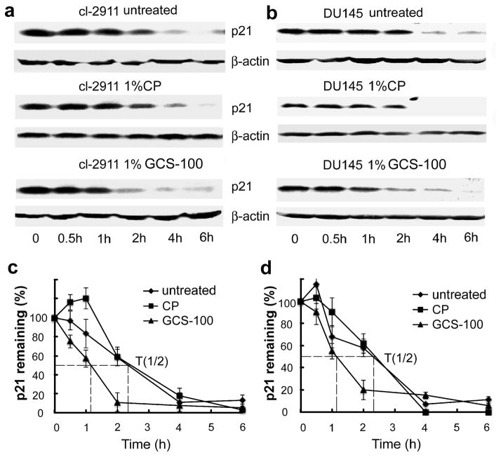
Figure 4.
The stabilization of p21 by Gal-3 is impaired by GCS-100. LNCaP Cl-2911 (a) and DU145 parental cells (b) were treated with 1% GCS-100 or 1%CP for 24 h, and treated with cycloheximide (50 μg/ml) for indicated times. Cells were harvested and subjected to Western blot analysis. Data are representative of three independent experiments. The graph of half-life of p21 in LNCaP cl-2911 (c) and DU145 cells (d) after GCS-100 treatment. p21 expression was quantified by densitometric analysis and normalized to β-actin. The relative abundance of p21 was represented as the percentage remaining relative to time zero.

