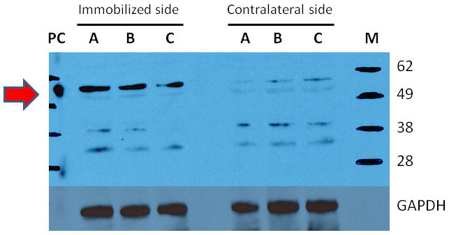
Figure 5. Protein expression of α7 acetylcholine receptors (α7AChRs) in muscle.

The tibialis muscles from immobilized and contralateral sides (N = 3 per side) previously harvested, were homogenized and subjected to immunoblots. Brain extract was used on positive control for α7AChRs, and glyceraldehyde 3-phosphate dehyrogenase (GAPDH) as the loading control. The letters M and PC indicate the molecular weight marker and positive control (Brain extract). The α7AChRs is located at 55kD molecular weight, indicated by the red arrow on the left. At 14 days after immobilization, the expression of α7AChRs was increased three fold compared to contralateral side.
