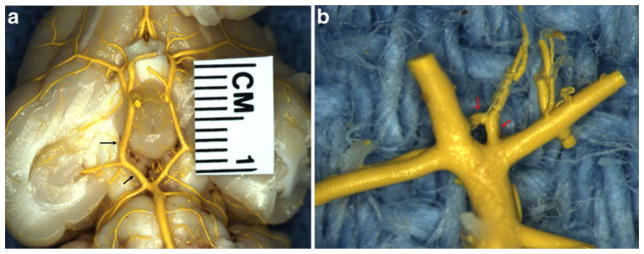
Fig. 2.

Gross photo of MICROFIL®-filled cerebral arteries harvested at 3 weeks after right common carotid ligation. a Shows the entire Circle of Willis. Note asymmetric enlargement of the right posterior communicating artery (arrow). b The high magnification of basilar terminus and its branches, showing prominent perforating arteries at the basilar terminus area (arrow)
