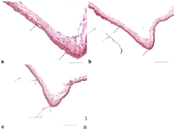
Fig. 4.
Serial sections from one 16-week subject illustrate artifact damage to the internal elastic lamella and endothelial cells (EC) during processing. a Showing local, sharp IEL and EC loss from processing (between two arrows) (H&E, original magnification ×400, bar=50 μm); b, c adjacent serial sections from the same subject as (a), showing the local IEL and EC loss (between two black arrows) at the same location in (a), as well the torn IEL and EC (dashed black arrows) (H&E, original magnification ×200, bar=100 μm)

