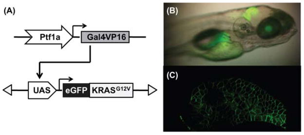
Fig. 1. Targeted expression of eGFP-KRASG12Vtransgene in zebrafish pancreas.

(A) Schematic drawing of the dyad Gal4/UAS system used to drive eGFP-KRASG12V transgene expression in the Ptf1a domain. (B) Lateral view under transmitted and fluorescent illumination of a larval fish at 5 days post fertilization (dpf), showing expression pattern of eGFP-KRASG12V transgene in the Ptf1a domain, including the retina, the hindbrain, and the exocrine pancreas. (C) Two-photon confocal image of the pancreas from a live 5 dpf larval fish, revealing the membrane localization of eGFP-KRASG12V protein. (For color version of this figure, the reader is referred to the web version of this book.)
