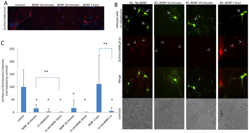
Figure 2. Decreased gephyrin expression with siRNA decreases surface GABAAR α1 on cultured amygdala neurons.
Panel A shows red-fluorescent surface GABAAR α1 subunits. The surface α1 subunits are decreased in neurons treated with BDNF for 10 or 20 minutes. Panel B shows single- and double-label ICC. GFP expression indicates neurons infected by LV-siGephyrin (arrows) and the surface GABAAR α1 is labeled with red fluorescence (arrowheads). B1 represents neurons infected with LV-siGephyrin and without BDNF treatment. The surface GABAAR α1 subunits were decreased only in infected neurons. B2 and B3 are neurons treated with BDNF for 10 or 20 minutes, respectively, and also infected with LV-siGephyrin. The surface α1 was decreased in infected neurons and non-infected neurons. B4 presents neurons treated with BDNF for 1 hour and infected with LV-siGephyrin. The surface α1 decreases only on infected neurons. Panel C illustrates the quantified surface α1 fluorescence intensity of Panel A and B. (LV-siG stands for LV-siGephyrin). (* P ≤ 0.01 comparing with the control. ** p ≤ 0.05 comparing between groups).

