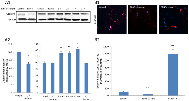
Figure 6. BDNF treatment leads to biphasic changes in gephyrin levels on cultured amygdala neurons.
Panel A1 is a result of a western blot for gephyrin from neurons treated with BDNF for 20 and 30 minutes and 1, 2, 4 or 17 Hours, respectively. A2 shows quantified western blot data demonstrating that gephyrin levels are decreased with 20 minutes of BDNF treatment. In a separate experiment, we showed that 1, 2 and 4 hours of BDNF treatment increased the gephyrin. (* P < 0.05 and ** p < 0.01, compared to control). Panel B1 shows ICC results of red fluorescent-labeled gephyrin on neurons exposed to BDNF for 20 minutes or 4 hours. B2 represents the quantified Gephyrin fluorescence intensities from ICC. (** p < 0.01 and *** p < 0.004, compared to control).

