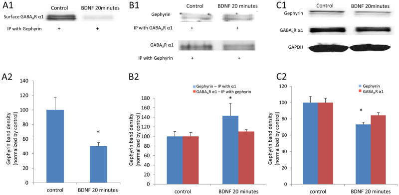
Figure 7. Decreased GABAAR α1 subunits on cell membranes were associated with gephyrin changes induced by rapid BDNF treatment.
Panel A1 is a western blot result of the surface GABAAR α1 subunits from biotinylated protein samples and then co-immunoprecipitated with an antibody against gephyrin. The surface GABAAR α1 specifically bound to gephyrin was significantly decreased by 20 minutes of BDNF treatment. (* p < 0.05, compared to control) (A2). Panel B1 shows co-IP results of gephyrin and GABAAR α1 from neurons treated with BDNF for 20 minutes and compared to untreated control neurons. B2 shows that gephyrin bound to GABAAR α1 was increased with BDNF treatment (* p < 0.05, compared to control). Panel C1 is a western blot result of gephyrin and total GABAAR α1 subunits. There is no change in total GABAAR α1 while gephyrin was significantly decreased (C2). (* p < 0.05, compared to control).

