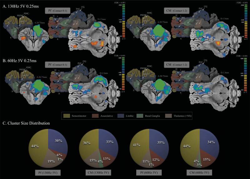
Figure 5.
fMRI BOLD effects with high amplitude (5V) stimulation (FDR<0.001). Both high frequency (A) and low frequency (B) stimulation evoked a similar negative BOLD response in the sensorimotor, associative, and limbic areas for Pf and CM DBS (C). The cluster distribution shows that regardless of stimulation frequency or DBS contact, the distribution remained stable (<5% variance). The average cluster size distribution percentage in 5V was: sensorimotor 41.25% ± 3.78; limbic 33% ± 2.16; associative 11.5% ± 3.87; BG 2.25% ± 1.5; and NS 12.25% ± 6.4. Abbreviations: NS, non-specific areas; CM, centromedian thalamic nucleus; CP, caudate and putamen; CT, central thalamic nucleus; DACC, dorsal anterior cingulate cortex; DPCC, dorsal posterior cingulate cortex; HP, hippocampus; IC, insular cortex; ITG, inferior temporal gyrus; PFC, prefrontal cortex; PHC, parahippocampal gyrus; PIMC, primary motor cortex; Pf, parafascicular thalamic nucleus; PP, pineal gland and perirhinal cortex; PPC, prepyriform cortex; PREMC, premotor cortex; PSS, primary sensory cortex; VAT, ventroanterior thalamic nucleus.

