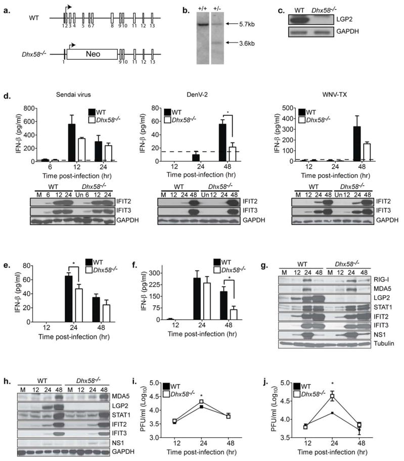
Figure 1. LGP2 is a positive regulator of RLR signaling in primary fibroblasts and myeloid-derived cells.
(A) LGP2 gene targeting strategy and predicted gene disruption. (B) Southern blot analysis of Dhx58+/+ and Dhx58+/− mice (WT allele = 5.7kb; Dhx58−/− allele = 3.7 kb). (C) Immunoblot analysis of mouse embryo fibroblasts (MEFs) from C57BL/6 (WT) and Dhx58−/− mice treated with type I IFN for 24 hours. (D) WT and Dhx58−/− MEFs were mock-infected (M) or infected with Sendai virus (200HA units/ml; left); DENV-2 (MOI 1.0; middle); and WNV-TX (MOI 1.0; right). IFN-β in the supernatant was measured by ELISA (upper) and interferon-stimulated gene (ISG) expression assessed by immunoblotting (lower) at the indicated hours post-infection. (E–I) Primary bone-marrow derived dendritic cells (DC) and macrophages (Mφ) recovered from WT and Dhx58−/− mice were mock-infected or infected with WNV-TX at an MOI of 1. Cells and culture media were harvested at the times indicated for determination of IFN-β production (DCs=E, Mφ=F), ISG expression (DCs=G, Mφ=H), and virus load (DCs=I, Mφ=J). Dotted line represents the limit of assay sensitivity. Graphs show the mean +/− standard deviation from triplicate samples from three independent experiments. Asterisks denote P < 0.05.

