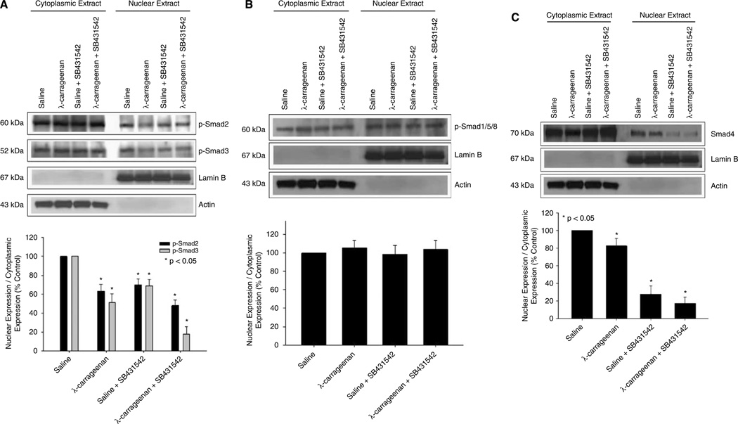
Figure 5.
Expression of Smad proteins in brain microvessels during CIP in the presence and absence of an ALK5 inhibitor. Western blot analysis of microvessels isolated from rats treated with saline or λ-carrageenan in the presence and absence of SB431542, a selective ALK5 inhibitor. Cytoplasmic and nuclear extracts from rat brain microvessels (20 µg) were resolved on a 10% sodium dodecyl sulfate–polyacrylamide gel and transferred to a PVDF membrane. Samples were analyzed for expression of p-Smad2 and p-Smad3 (A) as well as p-Smad1/5/8 (B) and Smad4 (C). P-Smad2 was detected using the polyclonal antibody Ser465/467 (1:500 dilution), p-Smad3 was detected using the polyclonal antibody Ser423/425 (1:500 dilution), pSmad1/5/8 was detected using the polyclonal antibody Ser463/465 (1:500 dilution), and Smad4 was detected using the polyclonal anti-Smad4 antibody (1:500 dilution). Relative levels of Smad proteins were determined by densitometric analysis and the nucleus/cytoplasm ratio was calculated. Results are expressed as mean ± s.d. of three separate experiments. Asterisks represent data points that are significantly different from control.

