
Figure 4.
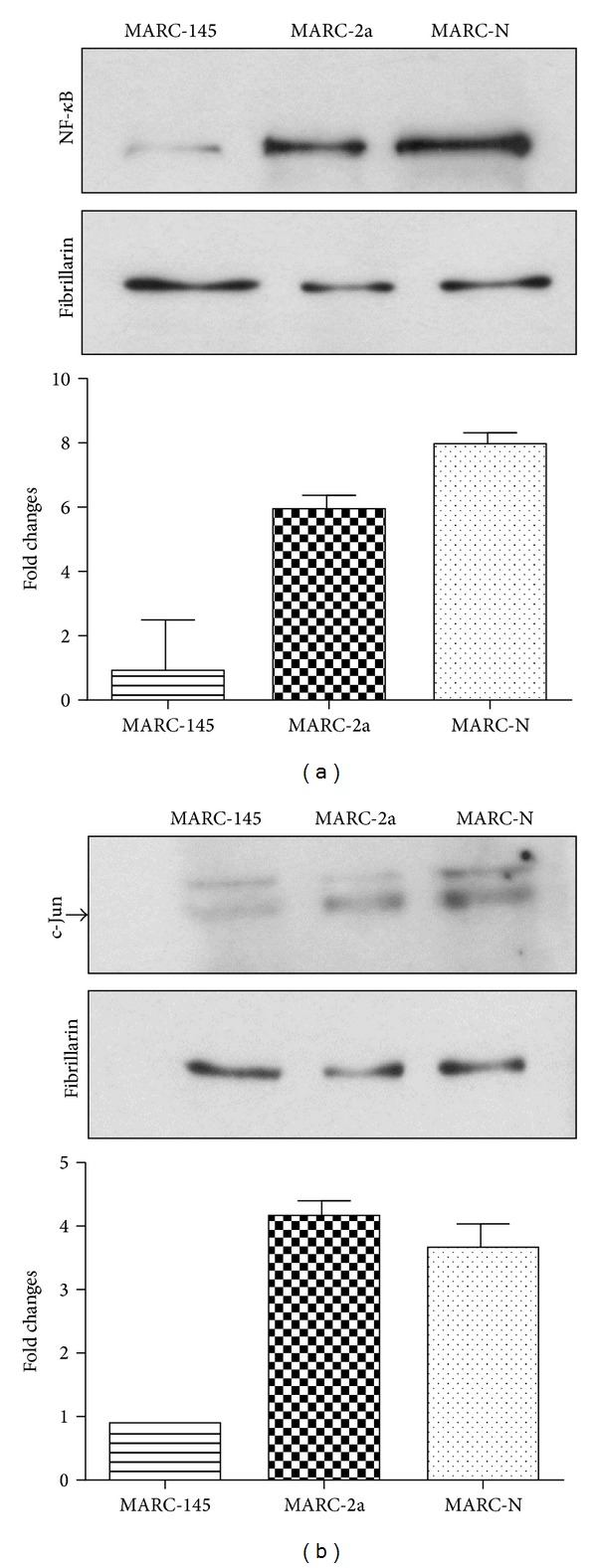
Western blot analysis of NF-κB and AP-1 activation. Equal amounts of nuclear protein fractions were separated on 10% SDS-PAGE. Cytoplasmic fraction contamination was evaluated by α-tubulin antibody (data not shown). Fibrillarin antibody was used as a nuclear marker and also as a loading control. Fold changes in protein amounts are plotted. Western blot with (a) NF-κB rabbit monoclonal antibody and (b) c-Jun mouse monoclonal antibody. Arrow shows the position of the 48 kDa band corresponding to the phosphorylated form of the c-Jun protein.