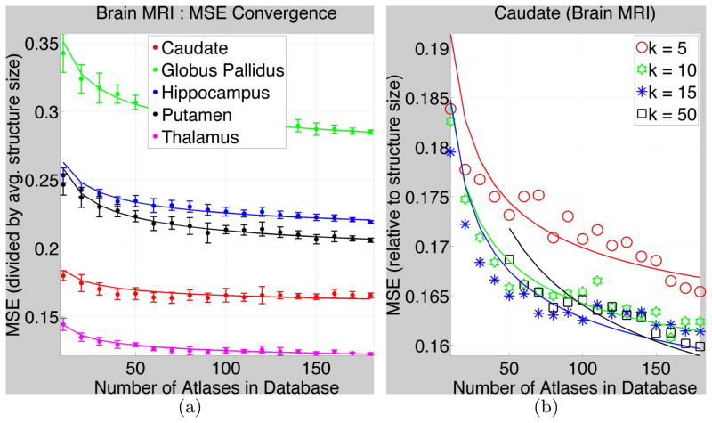
Fig. 1. MSE Convergence for Subcortical Structures in Brain MR images.
(a) The dots and the error bars show MSE(Mj) and the standard deviation, respectively, (divided by the average true size of structures in database) for k = 10. The parametric fitted curves are shown by solid lines. Table 1 gives the parameter values. (b) shows MSEs and fitted curves for the caudate (as an example) for varied k.

