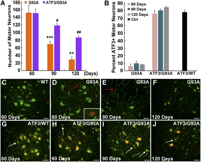
Fig. 1.
Forced ATF3 expression promotes motor neuron survival. (A) Lumbar ventral horn motor neurons immunostained for the neuronal marker NeuN with an area ≥ 450 μm2 were quantified in the SOD1G93A compared with ATF3/SOD1G93A mice at 60, 90, and 120 d. A significant difference in the number of surviving motor neurons was identified. Data are presented as mean ± SEM with n = 5 mice per group per time point. **P < 0.01; ***P < 0.001 by ANOVA with Bonferroni postanalysis. A significant difference in the number of motor neurons in ATF3/SOD1G93A at 90 and 120 d relative to 60 d was also detected. #P < 0.05; ##P < 0.001 by ANOVA with Bonferroni postanalysis. (B) A significant difference was found in the percentage of ATF3-positive motor neurons in ATF3/SOD1G93A compared with SOD1G93A mice at all time points (P < 0.0001 by ANOVA). No difference in the proportion of ATF3-positive motor neurons was detected within each group over time (P > 0.05 by ANOVA with Bonferroni postanalysis). ATF3 expression in control WT and ATF3/WT mice at age 120 d is presented. (C–J) Representative merged images of motor neurons double stained for NeuN (green) and ATF3 (red) (nonmerged images are presented in Fig. S1). (Scale bar: 50 μm.) (D and E) Some endogenous ATF3 induction is detected in the SOD1G93A mice (red arrows and Inset). (G–J) Motor neurons that do not express ATF3 in the ATF3/WT and ATF3/SOD1G93A mice are marked by a white arrow.

