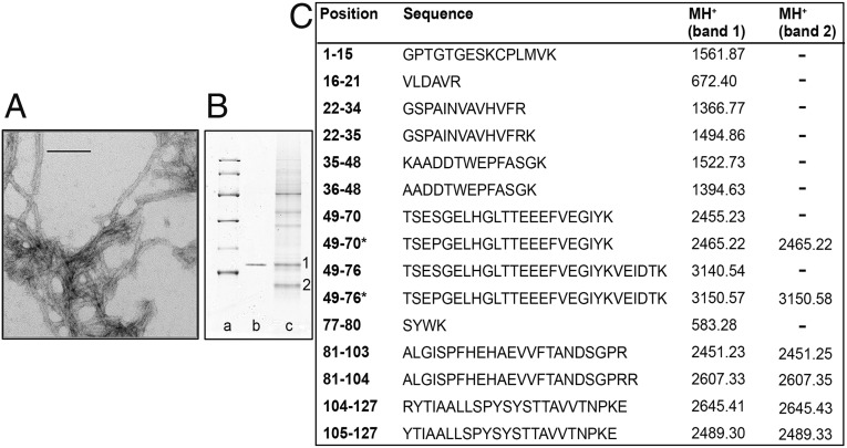
Fig. 1.
Ex vivo amyloid fibrils. (A) Transmission electron microscopy of amyloid fibrils extracted from spleen. (Direct magnification: 230,000×; scale bar, 100 nm.) (B) SDS 15% PAGE under reducing conditions. Lane a: marker proteins (14.4, 20.1, 30.0, 45.0, 66.0, and 97.0 kDa, respectively); lane b: 0.5 μg recombinant Ser52Pro TTR; lane c: 60 μg ex vivo amyloid fibrils from spleen. 1 and 2 indicate bands subjected to mass mapping analysis. (C) Tryptic peptides obtained by digestion of SDS/PAGE bands were analyzed by MALDI-MS and nano-LC MS-MS. MH+ monoisotopic values are reported for each peptide; asterisks indicate the presence of the Ser52Pro substitution. Cysteine is carbamidomethylated.

