Fig. 4.
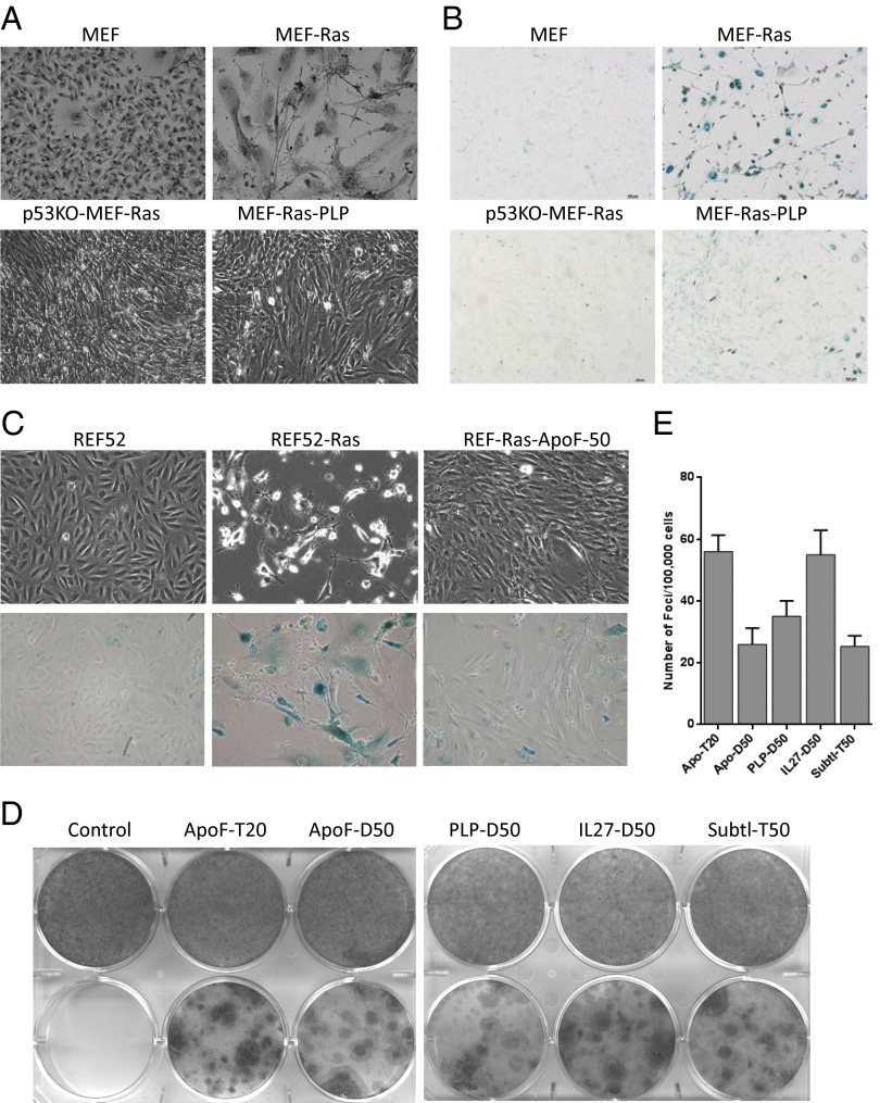
NASP expression allows MEF and REF52 cells to overcome Ras-induced senescence and undergo transformation. (A) Photographs taken under light microscope, and (B) β-galactosidase staining of primary MEFs that were transduced with the empty vector PLV-bleo (MEF), transduced with the lentiviral H-RasV12 expression construct PLV-rasV12-bleo (MEF-Ras), MEFs (p53−/−) transduced with PLV-rasV12-bleo (p53KO-MEF-Ras) or transduced with PLV-rasV12-bleo and a lentiviral NASP (PLP) expression construct (MEF-Ras-PLP). Transduced cells were selected in bleomycin, allowed to grow for 21 d, and then photographed/stained. Note transformation phenotype in cultures of p53−/− MEFs expressing Ras or in those coexpressing Ras with PLP, but not in those expressing Ras alone. (C) Phase contrast images (Upper) and β-galactosidase staining (Lower) of REF52 cells that were (in order from left to right) (i) transduced with the empty vector PLV-bleo, (ii) transduced with the lentiviral H-RasV12 expression construct PLV-rasV12-bleo, or (iii) transduced with PLV-rasV12-bleo and a lentiviral NASP (IL-27-T50) expression construct. Transduced cells were selected in bleomycin, allowed to grow for 18 d, and then photographed/stained. (D) Representative images of methylene blue staining of REF52 cells transduced with NAGE vector construct (control) or different NAGE expression constructs either with PLV-Bleo (Upper) or with PLV-rasV12-bleo (Lower). Tested NAGEs included those derived from apolipoprotein F (ApoF), pancreatic lipase-related protein-1 (PLP), interleukin-27 (IL27), and subtilisin/kexin type 9 (Subtl). Here and in E, the library from which the NAGE was isolated is indicated by D or T (for dimer or trimer) and/or the number of amino acid residues in the peptide inserts (20 or 50). (E) The frequency of foci formation was calculated from triplicate plates (one of which is shown in D).

