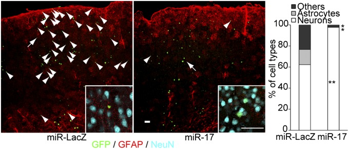
Fig. 5.
In vivo roles of miR-17/106 in developing mouse forebrains. Lentiviruses that permitted OE of miR-LacZ or miR-17 were microinjected in utero into the cerebral ventricle of mouse embryos at E10.5, and the fates of the infected cells were examined in the cerebral cortex at P30 by immunohistochemistry (n = 3). Neuronal nuclei were stained with an antibody against NeuN. Arrowheads indicate nonneuronal cells, which include GFAP+ astrocytes. Most GFAP-negative nonneuronal cells (defined as “Others” in the graph) appeared to be immature astrocytes. Higher magnification images of the cells indicated with arrows are shown as insets. (Scale bars, 50 μm.) *P < 0.05; **P < 0.01.

