Figure 1.

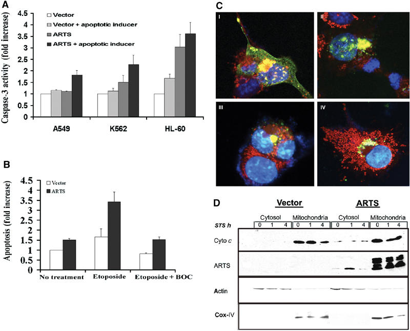
ARTS induces caspase-3 activity in response to a variety of apoptotic inducers, and caspase activity affects ARTS localization. Cells were transiently transfected with either control vector or the AU5-ARTS construct and treated with various apoptotic inducers. (A) ARTS induces caspase-3 activity in response to different apoptotic inducers. A549 cells were treated with TGF-β, K562 and HL-60 with ara-C. Caspase activity is presented as fold increase compared to cells transfected with control vector without apoptotic treatment. (B) COS-7 cells were treated with 50 μM etoposide for 16 h with and without BOC. (C) Immunofluorescence assay of COS-7 cells with MitoTracker® Red CMXRos and anti-ARTS antibodies. The cells were treated with 50 μM of etoposide for 16 h without (C I, II) and with BOC (III, IV). Cells treated with etoposide show typical morphological changes associated with apoptosis and accumulated ARTS in the nucleus (C I, II). Upon addition of caspase inhibitors nuclear translocation of ARTS is blocked, although perinuclear clustering of ARTS-positive mitochondria is still observed (C III, IV). (D) COS-7 cells transfected with AU5-ARTS or AU5-empty vector were treated with 1 μM STS for 0, 1 and 4 h prior to cell fractionation. Western blot analysis using anti-cytochrome c antibodies (upper panel) show cytochrome c restricted to the heavy membrane/mitochondrial fraction. Reduced cytochrome c levels were seen after 4 h of treatment with STS. Upon overexpression of ARTS, some cytochrome c was found in the cytosol. This is consistent with the increased levels of apoptosis seen under these conditions. The middle panel shows the distribution of ARTS in different cell fractions, using an anti-ARTS antibody (Sigma). In nonapoptotic cells, the vast majority of ARTS was found in the mitochondria. However, low amounts of ARTS protein were also detected in the cytosol of cells overexpressing ARTS. Upon apoptotic induction (1 h with STS), ARTS was released from the mitochondria and elevated levels were found in the cytosol. An antibody against the OxPhosComplex IV subunit IV (COX IV) was used as a mitochondrial marker.
