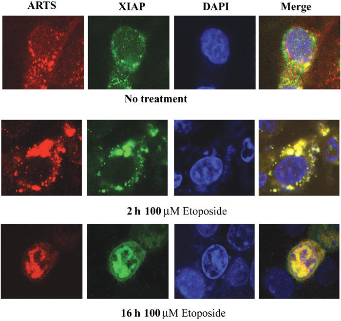
Figure 5.

ARTS and XIAP co-localize during apoptosis. Immunofluorescence assay of COS-7 cells with anti-ARTS antibodies (Red), and anti-XIAP antibodies (green). The nucleus is stained with Dapi (blue). Cells were treated with 100 μM etoposide for 2 h (mid panel) and for 16 h (lower panel), as compared to non-treated cells (upper panel). The cells were analyzed using confocal microscopy (× 60). The merged images (right panel) are shown in yellow. Nonapoptotic cells had very little overlap between ARTS and XIAP staining (top panel). In nonapoptotic cells, ARTS was primarily localized to mitochondria, whereas XIAP staining was cytoplasmic, with some perinuclear concentration. At 2 h after the induction of apoptosis, co-localization of ARTS and XIAP occurred mainly in the vicinity of the nucleus (mid panel). After 16 h of apoptotic induction, both proteins perfectly co-localized in the nucleus.
