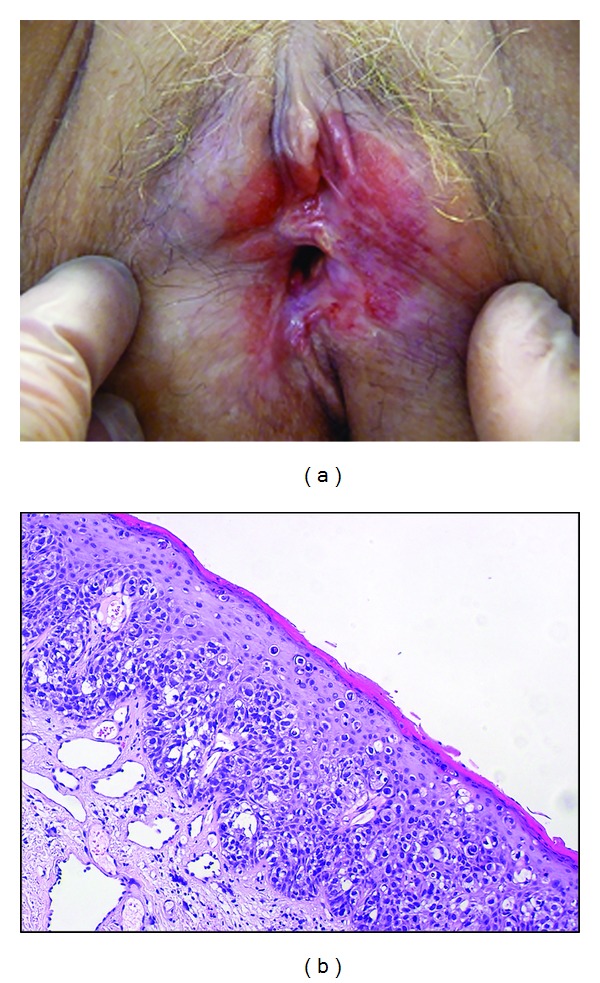
Figure 3.

(a) Wide red area extending mainly on the left side of the vulva in a 65-year-old woman with Paget's disease. (b) Representative example of Paget's disease biopsy specimen showing many glandular neoplastic cells with clear cytoplasm present singly or in small nests within the epidermis.
