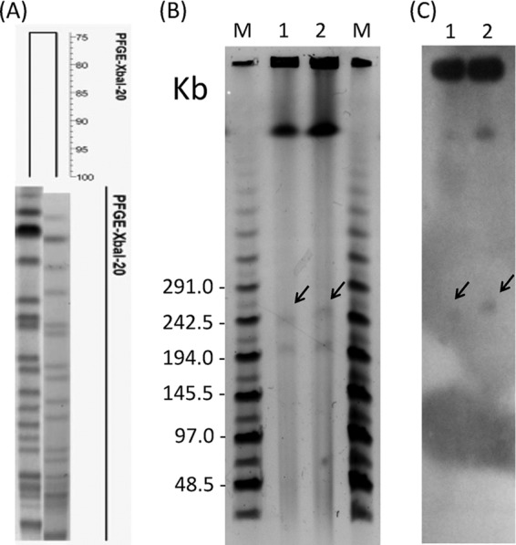
FIG 1.

PFGE analysis of R. planticola. (A) XbaI-digested chromosome fragments were separated by PFGE, and the dendrogram was produced by BioNumerics software. (B) S1-nuclease-digested plasmid profiles separated by PFGE. (C) S1-nuclease-digested plasmid profiles hybridized with a blaIMP-8 probe. Lanes M, MidRange II PFG marker; lanes 1, isolate 139; lanes 2, isolate 193. The arrows show the locations of blaIMP-8 genes.
