
Figure 4.
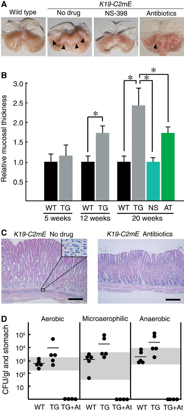
Suppression of gastric hyperplasia either by treatment with a COX-2 inhibitor (NS-398) or antibiotics. (A) Macroscopic photographs of the stomach at 20 weeks of age of the wild-type (left) and K19-C2mE (right) mice, with respective treatments as indicated. Arrowheads in ‘no-drug' indicate the hypertrophic lesion. In the antibiotics-treated stomach, a mild hypertrophic lesion is still found (arrowhead). (B) Relative mucosal thickness at 5, 12 and 20 weeks of age is presented as the mean±s.d. (i.e., the mean thickness divided by that of the wild type). WT, wild type; TG, nontreated K19-C2mE; NS and AT, K19-C2mE treated with NS-398 and antibiotics, respectively. *P<0.05. (C) Histopathology of the glandular stomach in the untreated (left) and antibiotics-treated (right) K19-C2mE mice, respectively (H&E staining). Inset in the left panel shows a higher magnification of the mononuclear cell-infiltrated submucosa. Bars, 300 μm. (D) Bacterial counts in the glandular stomach at 20 weeks of age. CFUs per mouse glandular stomach are indicated as closed circles. Horizontal bars indicate the mean CFU. Culture conditions are shown on top. WT, wild type; TG, K19-C2mE; TG+At, K19-C2mE with antibiotics. Gray horizontal zones indicate the ranges of bacterial counts in wild-type mice.