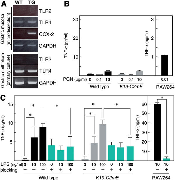
Figure 5.

Stimulations of TLR on the gastric epithelial cells with bacterial PGN or LPS. (A) RT–PCR for TLR2 and TLR4 mRNAs using total RNA from gastric mucosa sampled by LMD (top) and primary culture of the epithelial cells (bottom), respectively. COX-2 in LMD-sample RT–PCR was used as positive control. GAPDH was used as endogenous controls. WT, wild type; TG, K19-C2mE. Gastric epithelial cells were treated with PGN (B) and LPS (C) for 20 h. Concentrations of TNF-α in the supernatants are presented as the mean±s.d. RAW264 mouse monocyte cells were used for positive control. For TLR4 blocking, anti-TLR4 antibody was added at 10 μg/ml (blue bars). *P<0.05.
