FIG 3.

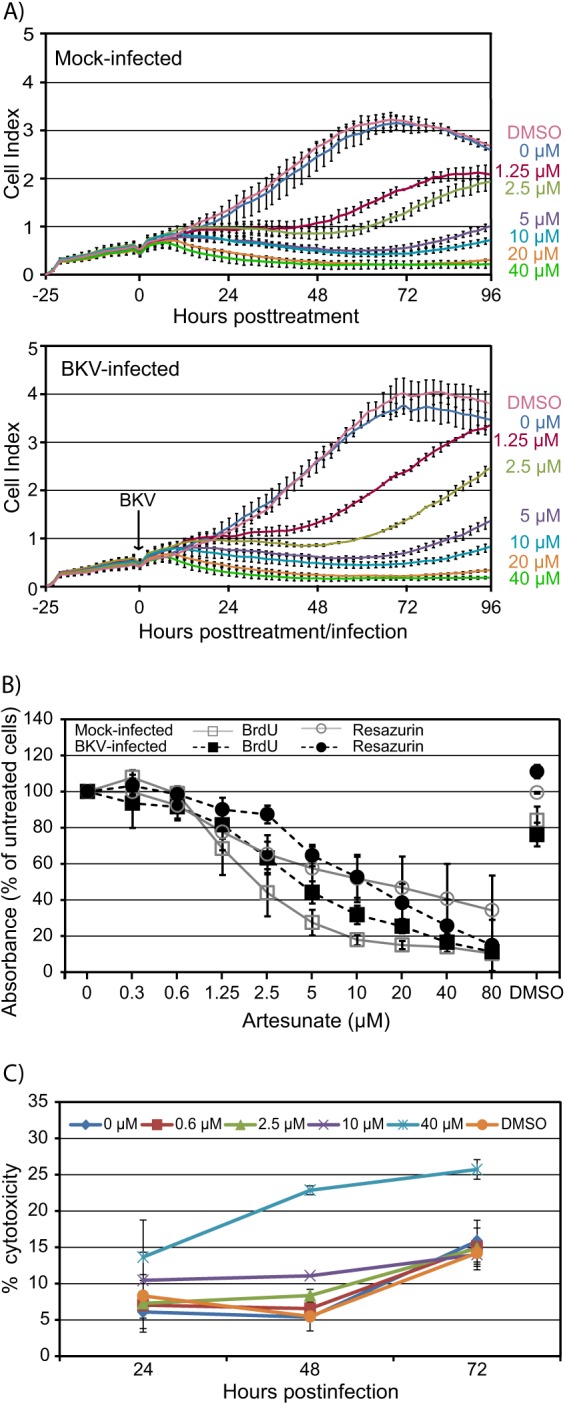
Effect of increasing concentrations of artesunate on viability of mock- and BKV-infected RPTECs. (A) Real-time cell proliferation. RPTECs were seeded in an E plate 16, and 25 h later, half of the medium in the wells was replaced by growth medium with artesunate to reach the indicated concentrations (top). In some wells, purified BKV Dunlop was also added (bottom). The CI, a combined measure of cell adhesion, proliferation, and size, was monitored from the time of seeding until 96 h posttreatment and infection by an xCELLigence RTCA DP instrument. The CIs, normalized to the value for the cell-free background, are shown as mean values ± SDs of 2 wells. The results of one representative experiment out of two are shown. (B) DNA replication and metabolic activity. Cellular DNA replication (BrdU) and the total metabolic activity (resazurin) of artesunate-treated mock- and BKV-infected RPTECs were measured at 72 hpi (i.e., 70 h posttreatment). Mean values of the percent absorbance of untreated cells ± SDs of three experiments (each experiment was performed in three wells) are presented. (C) Cytotoxicity. BKV-infected RPTECs were treated with the indicated concentrations of artesunate from 2 hpi. The LDH levels in the supernatant and the LDH levels in the well after complete lysis of the cell layer (total LDH) were measured at 24, 48, and 72 hpi. Cytotoxicity was calculated by dividing the amount of LDH in the supernatant by the total amount of LDH in the wells with the corresponding artesunate concentrations. Mean values of the percent cytotoxicity ± SDs from two to three experiments (each experiment was performed in three wells) are presented.
