Figure 4.

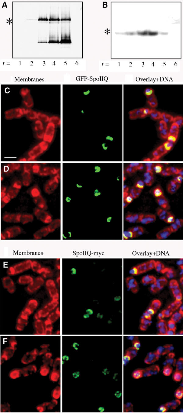
Western blot analysis and immunofluorescence of GFP-SpoIIQ and SpoIIQ-myc. (A) Immunoblot of GFP-SpoIIQ depicting specific proteolytic cleavage during engulfment. Full-length GFP-SpoIIQ is marked with an asterisk (*). (B) Western blot of SpoIIQ-myc shows no smaller degradation product. Full-length SpoIIQ-myc is marked with an asterisk (*). Immunofluorescence samples were taken at (C, E) t2 and (D, F) t3 and stained with Mitotracker Red (membranes in red), DAPI (DNA in blue) and SpoIIQ (GFP or Myc in green). (C, D) Localization patterns for GFP-SpoIIQ observed for fixed cells are the same as with live cells. (E, F) Localization of SpoIIQ-myc suggests that degradation occurs at the C-terminus of the protein since early patterns are similar to GFP-SpoIIQ, but little signal is observed in late sporangia. Scale bar, 2 μm.
