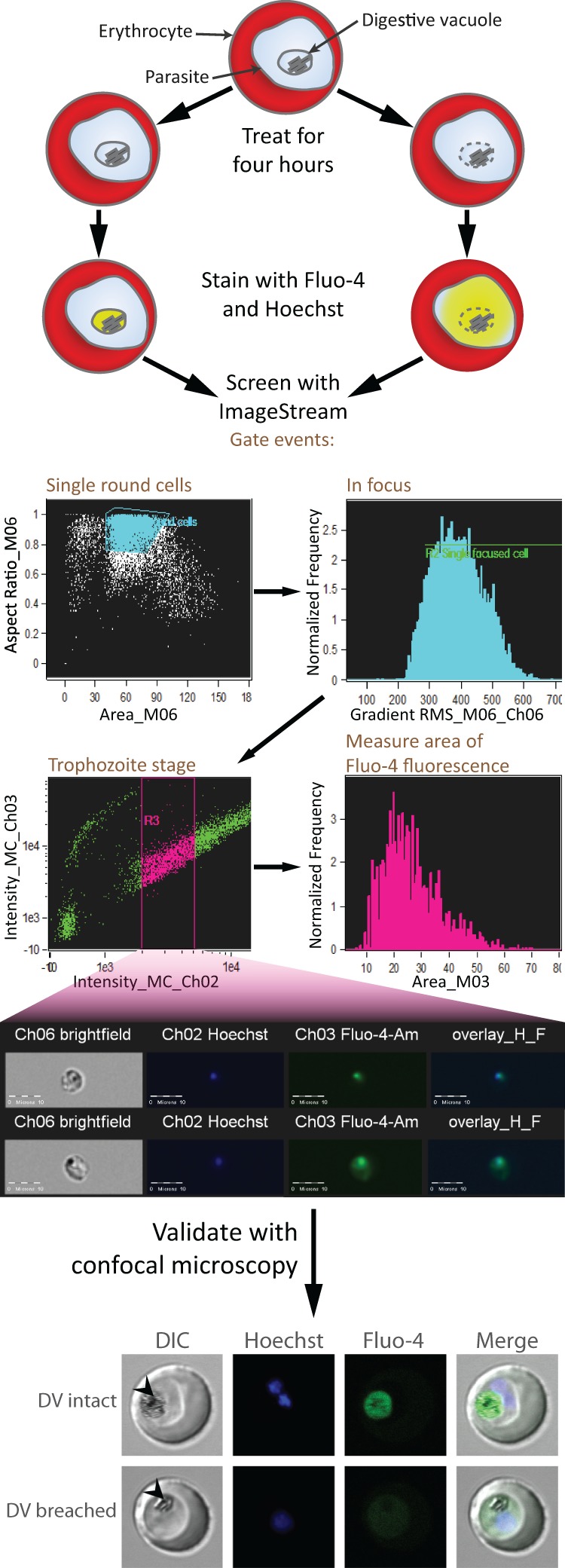
FIG 1.

Schematic representation of assay work flow. Erythrocytes infected with trophozoite-stage parasites are treated with the test compounds for 4 h. Following this, the cells are stained with Fluo-4-AM and Hoechst 33342 and analyzed with the ImageStream platform. Hits are validated by confocal microscopy. The confocal images shown are of representative trophozoites with intact or breached DV. Fluo-4 fluorescence localizes to the intact DV, whereas fluorescence is distributed to the parasite cytosol when the DV is permeabilized. Arrowheads indicate the DV. DIC, differential interference contrast.
