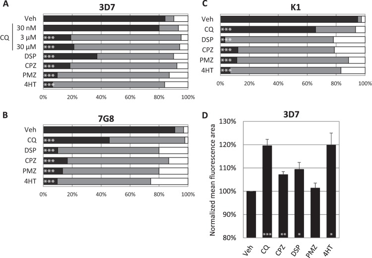
FIG 2.
Trophozoites were treated for 4 h with known lysosome destabilizers, stained with the Ca2+ probe Fluo-4-AM, and subsequently enumerated by confocal microscopy or analyzed with the ImageStream. Panels A, B, and C show the proportions of 3D7, 7G8, and K1 parasites with DV-localized fluorescence (black), cytosolic fluorescence (gray), and low or no fluorescence (white) after drug treatment. At least 30 infected erythrocytes were counted for each condition. Panel D shows the mean area of Fluo-4 fluorescence after treatment of 3D7. The mean areas for all conditions except PMZ were significantly different from the vehicle control (P < 0.05, n = ≥3). Data represent means ± the standard errors of the means. Veh, vehicle control. Concentrations: CPZ, 100 μM; DSP, 200 μM; PMZ, 200 μM; 4HT, 150 μM. When not stated otherwise, the CQ concentration was 3 μM. ***, P < 0.001; **, P < 0.01; *, P < 0.05.

