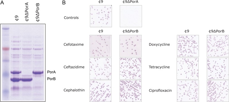
FIG 1.

Analysis of porin expression during MIC analysis. (A) Membrane proteins were isolated by Sarkosyl extraction, and 10 μg was separated on 8 to 12% bis-Tris acrylamide gels prior to Coomassie staining. Lane 1, ¢9; lane 2, ¢9ΔPorA; lane 3, ¢9ΔPorB. (B) Samples from the last well showing turbidity were plated on BHI agar and immunoblotted with the PorA-specific MAb MN14C11.6.
