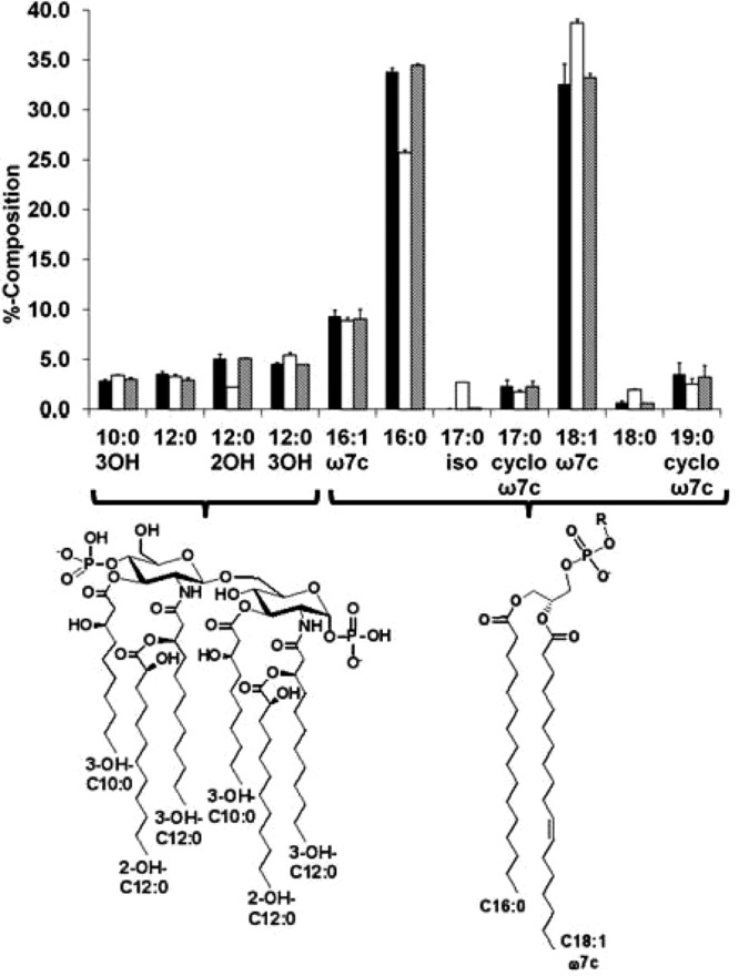
FIG 1.

Fatty acid composition of P. aeruginosa. The fatty acid content of each P. aeruginosa strain was determined by GC-FID analysis of the corresponding methyl ester. For each analysis, >95% of the summed peak areas on each chromatogram could be assigned. Values represent the averages of three independent samples isolated from cells grown on LB agar at 37°C for 18 to 24 h. Bars: ■, wild type; □, ΔfabY mutant; ▩, pfabY complemented mutant. (Bottom left) Hexa-acylated lipid A structure in P. aeruginosa with primary (3-OH-C10:0 and 3-OH-C12:0) and secondary (2-OH-C12:0) acylation; (bottom rght) P. aeruginosa glycerophospholipid substituted with the most common fatty acyl chains (C16:0 and C18:1 ω7c). Minor fatty acyl chains detected include acyl chains 16-carbons or longer that are saturated (i.e., C18:0), unsaturated (i.e., C16:1 ω7c), with a cyclopropane (i.e., C17:0 cyclo ω7c), or that are branched (i.e., C17:0 iso). The different phospholipid headgroups vary at position R.
