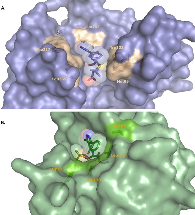
FIG 4.
Binding site for oxacillin in the K70D OXA-1/oxacillin complex (A) and the K84D OXA-24/40 complex (B). In panel A, the hydrophobic residues that line the binding pocket for the R1 side chain of oxacillin are drawn in gold and labeled. In panel B, hydrophobic residues are drawn in bright green and labeled. Oxacillin is shown as a stick representation surrounded by a transparent space-filling model.

