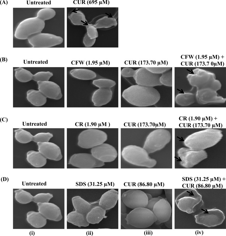
FIG 4.
Scanning electron micrographs showing corrugation of the cell surface, squeezing of the cell, and leakage of cytoplasmic content due to cell wall damage (arrows) in C. albicans after 24-h treatment with CUR and CWP agents at synergistic concentrations. (A) SEM of C. albicans treated or not with CUR (695 μM; 256 μg/ml) for 24 h showing corrugation of the cell wall and squeezing of cells. (B-i, C-i, and D-i) Controls; cells are intact and evenly shaped. (B-ii to -iv) Cells after treatment with 1.95 μM (1.79 μg/ml) CFW (B-ii), 173.7 μM (63.98 μg/ml) CUR (B-iii), and 1.95 μM (1.79 μg/ml) CFW plus 173.7 μM (63.98 μg/ml) CUR (B-iv). (C-ii to -iv) Cells after incubation with 1.90 μM (1.36 μg/ml) CR (C-ii), 173.7 μM (63.98 μg/ml) CUR (C-iii), and 1.90 μM (1.36 μg/ml) CR plus 173.7 μM (63.98 μg/ml) CUR (C-iv). (D-ii to -iv) Cells during incubation with 31.25 μM SDS (D-ii), 86.8 μM CUR (D-iii), and 31.25 μM (9 μg/ml) SDS plus 86.8 μM (31.97 μg/ml) CUR (C-iv).

