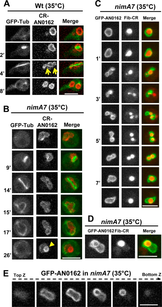
FIG 3.

Defective NE dynamics and nucleolar segregation in cells with partial NIMA function. (A and B) Mitotic dynamics of the NE marker protein CR-AN0162 and GFP-Tub in WT (A) (strain MG224) and nimA7 (B) (strain MG227) cells. The arrows in panel A indicate the normal NE restriction points during telophase. The arrowhead in panel B at 26 min shows the formation of the “theta”-shaped nuclear structure (see the text for details). (C) Mitotic dynamics of the nucleolus marked by fibrillarin-CR and GFP-AN162 in a nimA7 nucleus (strain MG294) that fails to generate two daughter nuclei after first mitosis even though the nucleolus first appears to be segregated into two. (D) The nucleolar protein fibrillarin-CR (strain MG294) is apparently enveloped by GFP-AN0162 (arrow). (E) Z sections through a nimA7 nucleus marked with GFP-AN162 reveal an NE protrusion and the abnormal shape of the nucleus (see also Movie S1 in the supplemental material). Bars, 5 μm.
