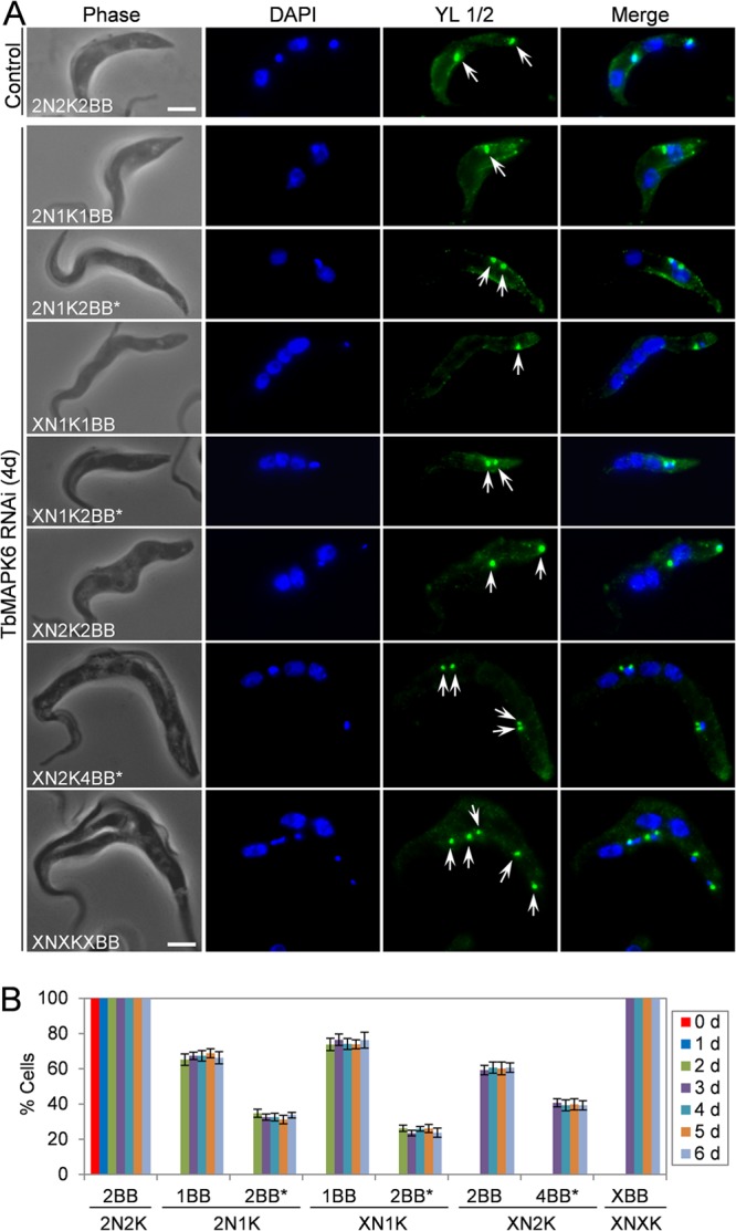
FIG 4.

Effects of TbMAPK6 RNAi on basal body duplication and segregation in the procyclic form. (A) Immunofluorescence microscopic analysis of the basal body in uninduced control and TbMAPK6 RNAi cells. The cells were immunostained with the YL 1/2 antibody and counterstained with DAPI for nuclear and kinetoplast DNA. The arrows indicate the mature basal bodies labeled by YL 1/2. The asterisks indicate closely associated basal bodies. Bars, 2 μm. (B) Percentages of TbMAPK6 RNAi cells with different numbers of basal bodies. The data are presented as the mean percentages ± SD of 200 cells counted for each time point from three independent experiments. Note that no 2N1K, XN1K, XN2K, and XNXK cells were detected at day 0 of TbMAPK6 RNAi. X > 2.
