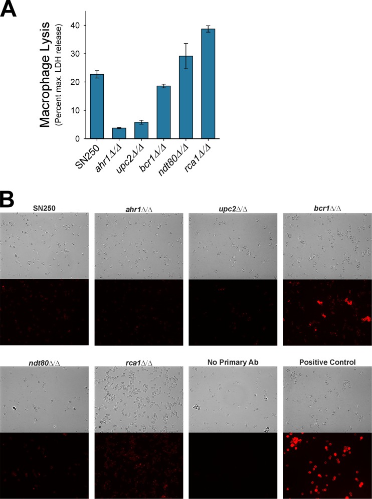
FIG 7.
C. albicans-induced pyroptosis does not correlate with surface 1,3-β-glucan exposure. (A) Macrophage lysis data are from the work of Wellington et al. (15) and are included in this figure for comparison to panel B. J774 macrophages were prestimulated with LPS for 2 h and then exposed to C. albicans WT (SN250) or mutants at an MOI of 2:1 for 5 h. LDH was measured from the supernatant of the cocultures as described for Fig. 1. (B) Photomicrographs of bright-field or Texas Red immunofluorescence of live C. albicans WT (SN250) or the indicated mutants incubated with a mouse monoclonal anti-1,3-β-glucan antibody followed by Texas Red-conjugated goat anti-mouse secondary antibody. All images were taken with the same exposure settings and processed only to adjust contrast. All images were processed identically.

