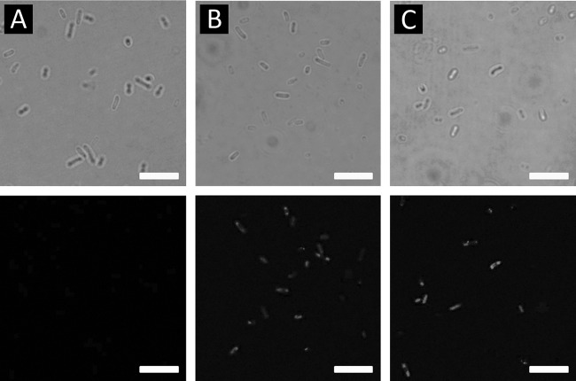
FIG 3.
Immunofluorescence analysis using differential interference micrographs (upper row) and immunofluorescence micrographs (lower row) of XL10-Gold cells (A) and XL10-Gold cells harboring pOW19F-PerPA3 (B) or pOW19F-PerPA4 (C). Cells were incubated with mouse anti-His probe antibody, followed by probing with goat anti-mouse IgG-FITC conjugate. Scale bars, 5 μm.

