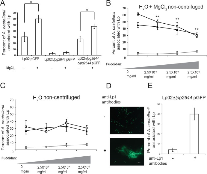
FIG 7.
Lcl-dependent autoaggregation potentiates the internalization of L. pneumophila in A. castellanii. (A) Infection of A. castellanii by Lp02/pGFP, Lp02 Δlpg2644/pGFP, and Lp02 Δlpg2644 clpg2644/pGFP in the absence or presence of 500 μM MgCl2 as measured by flow cytometry. (B and C) Infection of A. castellanii as measured by flow cytometry in the presence of 2.5 × 10⁻³ to 2.5 × 10⁻⁵ mg/ml fucoidan in deionized water with the addition of 500 μM MgCl2 (B) and in deionized water alone (C) with Lp02/pGFP (white squares), Lp02 Δlpg2644/pGFP (gray circles), and Lp02 Δlpg2644 clpg2644/pGFP (black triangles). (D) Fluorescence microscopy of Lp02 Δlpg2644/pGFP in the absence or presence of anti-Legionella pneumophila serogroup1 (anti-Lp1) agglutinating antibodies (1:1,000). (E) Infection of A. castellanii with isolated planktonic Lp02 Δlpg2644/pGFP and artificially aggregated Lp02 Δlpg2644/pGFP (without and with anti-Lp1 antibodies, respectively). Scale bar, 10 μm. * and **, statistically significant differences between the indicated strains (A) and compared to assays with 0 mg/ml fucoidan (B), respectively, by the two-tailed Student t test (P ≤ 0.01).

