
FIG 2.
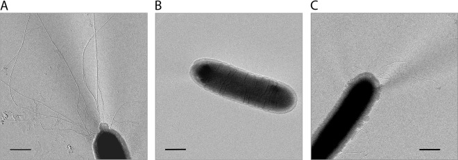
Electron micrographs of a representative sample of the T. thermophilus HB27 wild type (A), the ΔpilF deletion mutant (B), and the pilF::kat insertion mutant (C). Electron microscopic investigations were conducted by shadowing the cells with platinum-carbon. A total of 250 to 300 cells of each strain were analyzed. Bars = 500 nm.