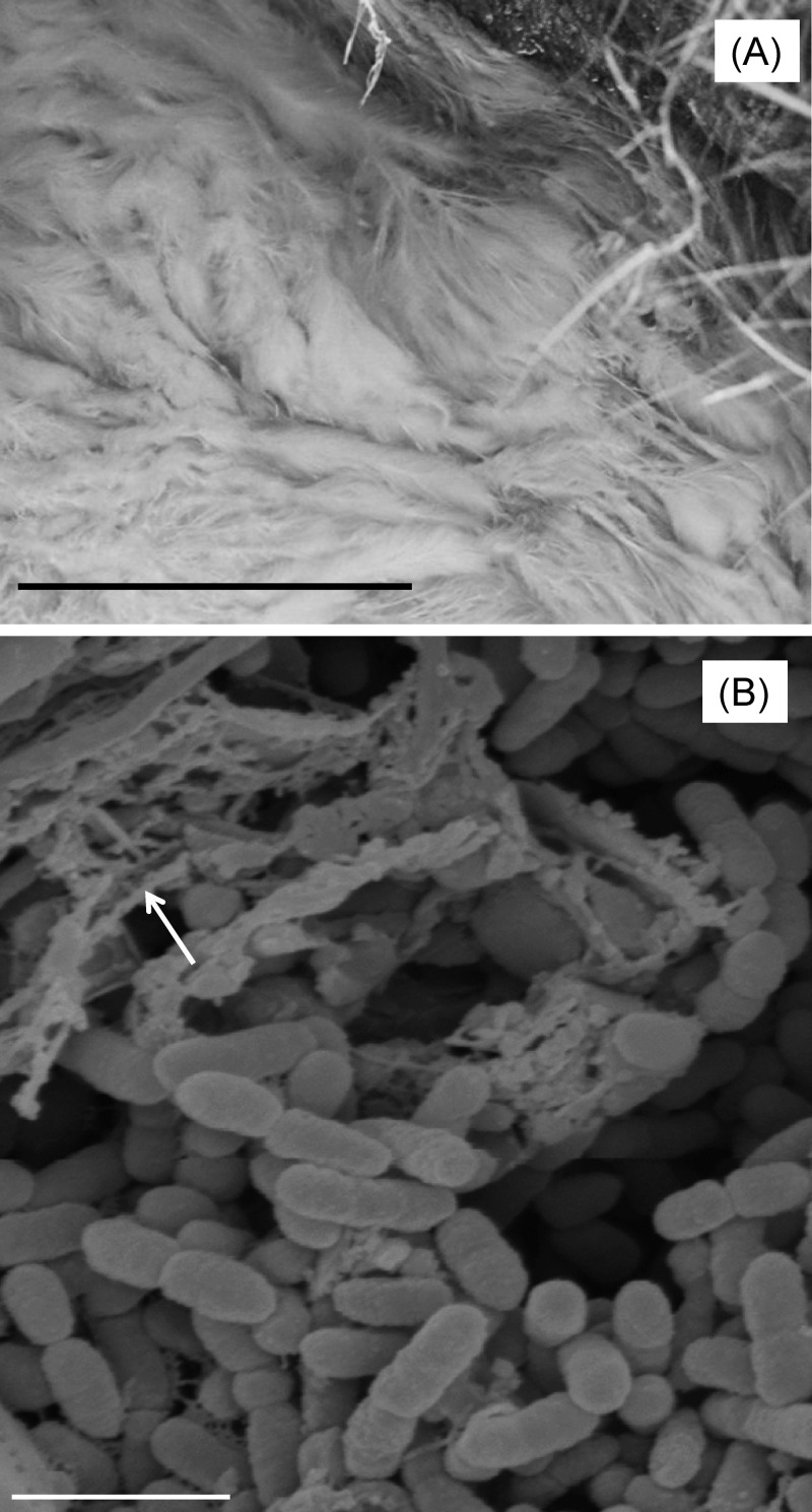
FIG 1.
(A) Acid streamers at Mynydd Parys, which served as the source of “F. myxofaciens” P3G (bar, 50 cm). (B) Scanning electron micrograph of “F. myxofaciens” P3G, showing aggregates of cells and dehydrated EPS (shown with an arrow) (bar, 2 μm). The sample was fixed in glutaraldehyde, critical point dried in liquid CO2, and viewed in an Hitachi S-120 scanning electron microscope.

