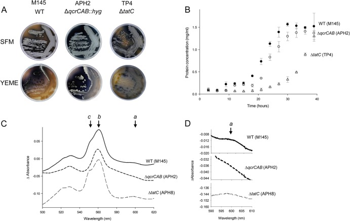
FIG 2.
Comparison of the S. coelicolor M145 and ΔqcrCAB and ΔtatC mutant strains. (A) Growth of S. coelicolor M145 (WT), APH2 (ΔqcrCAB), and TP4 (ΔtatC) on SFM or YEME agar plates. Spores of each strain were streaked and incubated at 30°C for 14 days. (B) Growth in liquid culture of the same strains used in the experiments whose results are shown in panel A. An amount of 108 spores of each strain, in triplicate, was inoculated into 100 ml TSB and grown at 30°C for 40 h with shaking at 200 rpm. Every 3 h, 1-ml samples were pelleted, cytosolic proteins released by boiling in 1 M NaOH for 10 min, and the protein content of each sample measured. (C) Absorption difference spectra of DDM-solubilized membranes (each at a protein concentration of 1.25 mg/ml) of strains M145 (WT), APH2 (ΔqcrCAB), and APH8 (ΔtatC ϕC31 [PhrdB-qcrCABHis10]). Spectra were initially collected under air oxidation, and then samples were reduced by the addition of dithionite. Note that the relevant genotype for each strain is given in the figure. (D) Expanded view of the 590- to 610-nm region of the same absorption difference spectra as shown in panel C.

