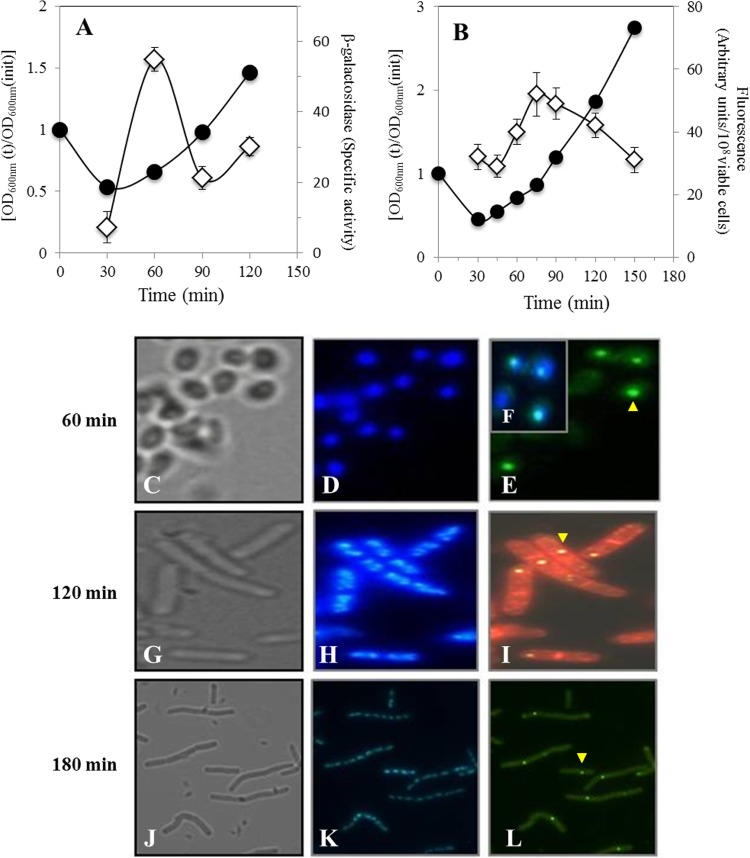
FIG 4.
Levels of β-galactosidase from a B. subtilis strain containing a disA-lacZ fusion (A), fluorescence emitted (B), and analysis of DisA-GFP synthesis (C to L) by a B. subtilis strain harboring a disA-gfp fusion during spore germination and outgrowth. Heat-shocked spores of B. subtilis PERM733 (disA-lacZ) and PERM1008 (disA-gfp) were germinated in 2× SG medium. (A). Samples collected at the indicated times were washed with 0.1 M Tris-HCl (pH 7.5), and cell pellets were stored at −20°C. Cell samples were processed for determination of β-galactosidase and for protein quantification as described in Materials and Methods. ●, OD600; ◊, β-galactosidase. (B) Samples collected at the indicated times were washed and suspended in PBS, and the samples' GFP fluorescence was measured as described in Materials and Methods. ●, OD600; ◊, GFP fluorescence. (C to L) Samples collected at 60 min (C to E), 120 min (G to I), and 180 min (J to L) were processed and photographed as described in Materials and Methods. (C, G, and J) Bright field; (D, H, and K) DAPI staining; (E, I, and L) GFP channel; (F) overlay of images in panels D and E. The sample in panel I (120 min) was stained with FM4-64 (red). The yellow arrowheads point to DisA-GFP.

