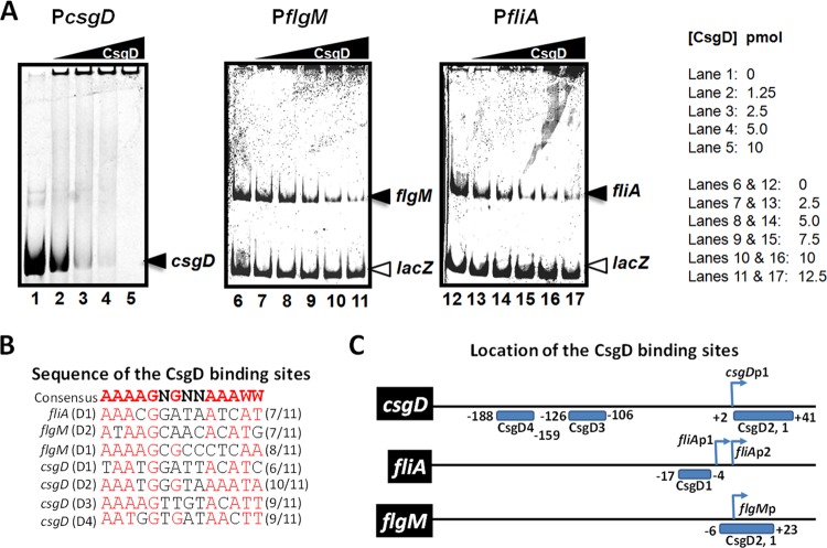
FIG 5.
CsgD binds the promoter region of the flagellar class 2 genes. (A) Gel shift assay of CsgD binding to its own promoter and the promoters of fliA and flgM. The protein was present at 1.25 to 12.5 pmol for 0.5 pmol of labeled DNA in a final volume of 20 μl. The concentration of CsgD is indicated by the height of the triangle above the gel, and the concentration of CsgD (in picomoles) in each lane is given to the right of the gels. (B) Consensus sequence for CsgD binding and sequences of the binding sites found around the −10 promoter element of the csgD, fliA, and flgM promoters. Individual DNA binding sites recognized by CsgD are labelled D1 to D4, which refers to specific positions within the target promoters. (C) Locations of the CsgD binding sites according to the transcription start site.

