
FIG 3.
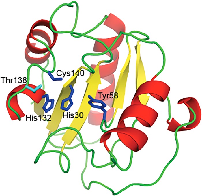
Structural model of Ph2119 endolysin. The model was built based on the crystal structure of D. melanogaster peptidoglycan recognition protein LB (PDB ID 1SK4). His30, Tyr58, His132, and Cys140, represented as blue sticks, are involved in Zn2+ binding. The secondary-structure elements alpha-helices, beta-strands, and loops are shown in red, yellow, and green, respectively.