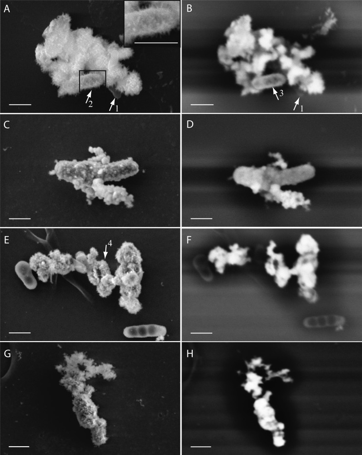
FIG 3.
SEM images of four nitrate-reducing strains cultured in the presence of 10 mM nitrate, 5 mM acetate, and ∼8 mM Fe(II). SE (5 kV) (left) and BSE (10 to 12 kV) (right) images of samples of bacterial strains Acidovorax strain BoFeN1 (A and B), Pseudogulbenkiania strain 2002 (C and D), Paracoccus denitrificans ATCC 19367 (E and F), and Paracoccus denitrificans Pd 1222 (G and H) are shown. Arrows 1 point to a nonencrusted cell of strain BoFeN1. Arrow 2 points to needle-like minerals. Arrow 3 points to the encrusted periplasm of a cell. Arrow 4 points to a complete encrusted cell. Bars, 500 nm.

